Stem cell therapy case study
 A lovely female Border Collie visited our clinic at around 1 year of age with sudden onset lameness in her right forelimb (RF). Her owner reported that this occurred during a walk the day before, and she had originally been non weight-bearing on this leg. On clinical examination the following day, she was 4/10 lame on her RF with pain on shoulder palpation.
Diagnosing forelimb lameness
– X-rays were taken with and without contrast and showed a region of biceps brachii tendon that had taken up some of the dye.
– A joint fluid sample showed significantly more joint fluid than would be expected in a normal joint, and it was tinged with blood.
– No marks or bruising were visible on her skin.
Shoulder injuries can be tricky to diagnose, as a tendon or ligament is often affected and can be difficult to identify on X-rays which are better at showing changes to the bones. This is why we used contrast, which involved injecting a special dye into the joint, as it helps the soft tissue structures show up more clearly on X-ray. In this case, the contrast X-ray was sufficient, but sometimes we have to perform more advanced imaging such as CT scans or MRI.
What can cause biceps tendon injuries in dogs?
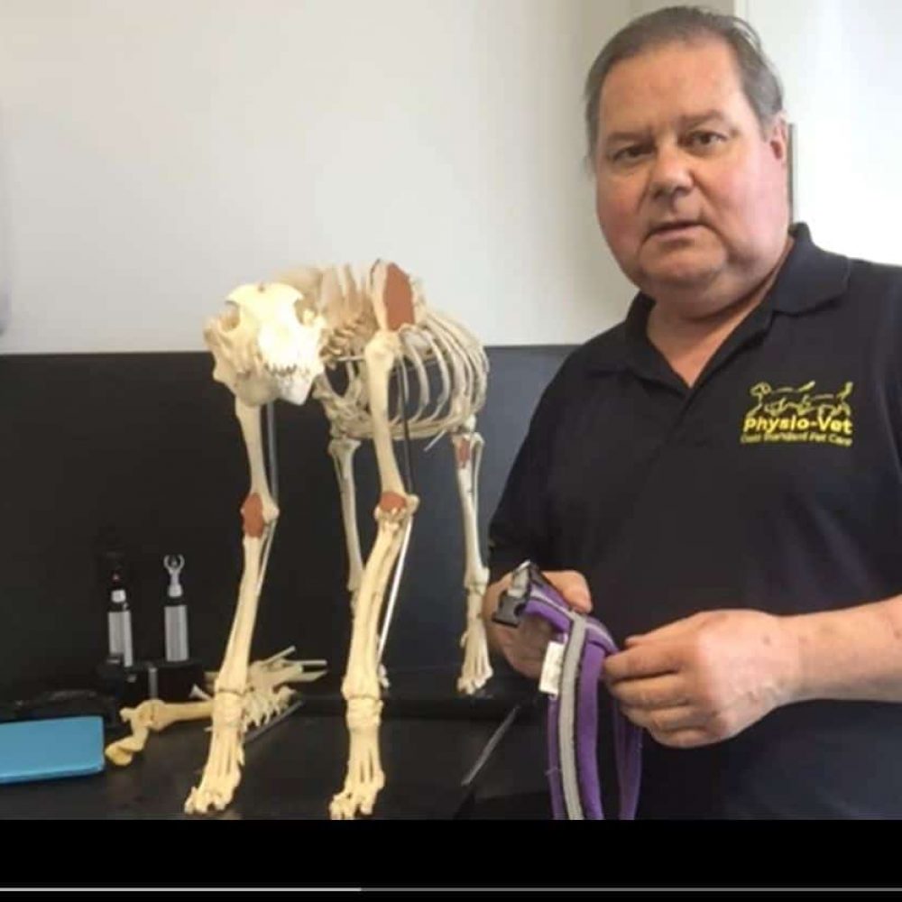
Lead vet, David, suspects that an ill-fitted harness is a likely cause of this case of lameness. This is because the chest straps and buckles crossed her biceps tendon putting pressure on it. We have put together two handy videos which demonstrate how to choose a harness that fits your pet:
How is a biceps tendon injury treated?
Our patient was put on strict rest, with a carefully controlled exercise plan according to her progress at follow-up appointments. She also received ongoing treatments of laser therapy and pulsed electromagnetic field therapy. However, a key component of her treatment plan was stem cell therapy.
What is stem cell therapy?
At Physio-Vet our team are passionate about keeping abreast of the latest cutting-edge therapies, and we have seen excellent results from stem cell treatments. A sample of bone marrow is taken from the patient’s humerus and processed in a centrifuge – this is a machine that spins at high speeds to create centrifugal forces. This separates out liquids of different densities into different layers. In this case, we wanted to separate out the stem cells from the other components of the bone marrow. These stem cells were then injected into the affected RF shoulder joint to help stimulate tendon repair.
Tendon injuries are often very slow to repair, but this patient’s lameness had resolved within a few weeks of receiving her stem cell therapy and we are delighted with her progress.
-
Previous
-
Next


